What is Anatomy Learning By Dr. Blanco Apps?
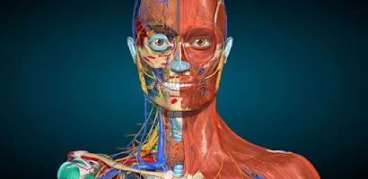
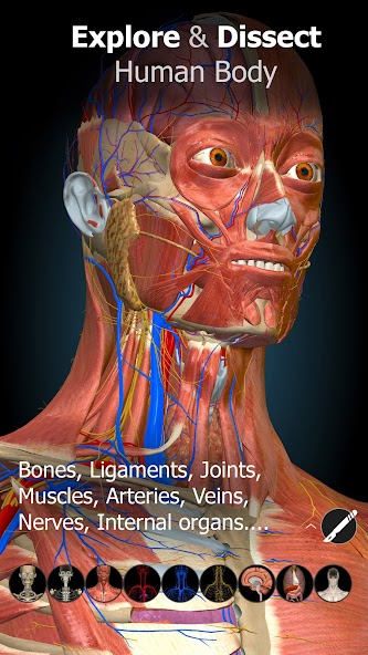
Anatomy Learning - 3D Anatomy is an interactive educational application designed to provide comprehensive three-dimensional models of human anatomy. It presents organs, bones, muscles, nerves, and vascular systems with detailed visualizations that can be rotated, zoomed, and dissected layer by layer. The platform supports annotation and labeling tools that allow users to identify structures and explore relationships between anatomical components. Its intuitive interface facilitates exploration by learners with varying levels of expertise, from secondary school students to advanced healthcare professionals. The realistic rendering techniques used in the models enhance depth perception and spatial understanding, making it easier to grasp anatomical complexity and topography. Interactive quizzes and learning modules help reinforce retention, offering customizable study paths that adapt to individual needs. Educational institutions and self-directed learners benefit from the modular organization of content, where systems are grouped logically and cross-referenced for integrated study. The program includes search capabilities that accelerate locating specific structures and provides layered views enabling progressive learning from superficial to deep anatomy. Users can create bookmarks and personal notes tied to particular views, aiding revision and long term study planning. Multimedia elements such as clinical case examples, functional animations, and comparative anatomy views enrich the learning context by linking structure to function. A focus on accessibility is evident through adjustable display settings, labeled languages, and scalable model fidelity suitable for a range of hardware configurations. Performance optimization ensures smooth interaction across devices while preserving model detail. Overall, the application functions as a versatile anatomy atlas that supports visual learning, practical review, and clinical correlation in a structured, approachable format. Learners can compare normal and pathological anatomy, measure distances, simulate surgical approaches, and export images for study materials, making the application suitable for classroom demonstrations, independent revision, and interdisciplinary collaboration among students, educators, and clinicians across multiple learning contexts.
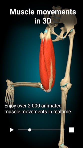
From an educational pedagogy perspective, Anatomy Learning - 3D Anatomy integrates multimedia pedagogy principles to promote active learning, spatial cognition, and cognitive load management. The program's layered dissection tools allow learners to control information pacing, exposing anatomical complexity incrementally to avoid overwhelming novices. Interactive labeling, quiz prompts, and timed challenges embed retrieval practice strategies that strengthen long-term memory consolidation. Visual learners gain considerable advantage from high-fidelity 3D models that link morphological detail with physiological function through animated sequences demonstrating muscle contraction, blood flow, and organ mechanics. Collaborative learning options allow groups to examine models synchronously, discuss findings, and annotate shared views for joint problem-solving exercises. Educators can design structured modules that align with curriculum objectives, sequencing topics from gross anatomy to system-level integration and clinical application. Formative assessment tools within the platform provide immediate feedback, guiding learners to remediate misconceptions and revisit targeted structures. The ability to simulate clinical scenarios or correlate anatomical features with common pathologies enhances transfer of learning to real-world applications. For students preparing for practical examinations, the interactive environment supports spatial recall and identification drills that mirror cadaver lab experiences without material constraints. Adaptive difficulty settings accommodate diverse proficiency levels, offering scaffolded learning paths that challenge advanced users while supporting beginners. The application also fosters interdisciplinary learning by linking anatomy to physiology, radiology, and basic surgical principles through layered content and integrated multimedia. As a supplement to traditional resources, this digital atlas reduces reliance on static diagrams by offering manipulable three-dimensional representations that can be studied repeatedly, paused, or reoriented. Overall, the instructional design emphasizes active engagement, immediate feedback, and contextualized practice to promote deeper conceptual understanding and improved retention among learners across educational stages. Students report increased confidence in identifying structures and relating anatomy to clinical signs through repeated, self-paced interactions with the models regularly.
Technically, Anatomy Learning - 3D Anatomy leverages modern graphics pipelines and optimized 3D asset management to deliver detailed anatomical models with responsive interaction. Models are constructed from polygonal meshes with layered textures and normal maps, augmented by skeletal rigs and morph targets where animations are required. A scene graph architecture allows hierarchical control of anatomical subsystems, enabling selective visibility, inverse kinematics for joint articulation, and efficient occlusion culling. The application uses progressive level-of-detail (LOD) techniques to adjust mesh complexity dynamically based on camera distance, balancing visual fidelity and runtime performance. High-resolution textures are streamed or tiled to minimize memory footprint while preserving surface detail when zoomed. Real-time lighting and shading models, including physically based rendering (PBR) materials, provide realistic tissue appearance under different lighting conditions, enhancing depth cues and material differentiation. Interaction interfaces are implemented with responsive gesture recognition and precision controls for rotation, translation, and cross-sectional slicing. Annotation data are stored in structured formats allowing export and import of labeled sets for study or teaching reuse. Measurements and volumetric assessments use built-in calibration and unit systems that support metric and imperial preferences for distance and volume calculations. Networked collaboration modes synchronize view states and annotations between participants through efficient serialization and delta updates, minimizing bandwidth. The software architecture separates presentation, model, and data layers to facilitate extensibility, plugin support, and integration with external educational systems. Security considerations are addressed by sandboxing asset loading and validating external resources to prevent malformed inputs. Performance profiling and automated testing routines help maintain stable frame rates and functional correctness across a variety of hardware profiles. Collectively, these technical choices create a robust platform that balances realism, responsiveness, and flexibility for diverse educational and professional use. Ongoing asset refinements and modular updates help keep the anatomical content current and academically relevant for medical education.
In terms of user experience, Anatomy Learning - 3D Anatomy emphasizes intuitive controls, clear visual language, and customizable study workflows to accommodate varied learner preferences. The main viewing canvas provides multi-touch or pointer-based navigation with smooth inertia and precise snapping options for aligning anatomical planes. Tool palettes offer quick access to system toggles, measurement tools, and annotation utilities while maintaining unobtrusive screen real estate for immersive study. Color-coding conventions and selective highlighting make it easy to differentiate tissues and trace pathways like neurovascular bundles or lymphatic channels. Custom playlists of views and annotated sequences can be assembled to create guided tours for specific lessons or revision sessions. Accessibility features include adjustable font sizes for labels, color contrast options for visibility, and alternate labeling modes that reduce clutter by showing only relevant terms. For users who prefer a checklist-driven approach, progressive reveal modes can present structures incrementally and lock completed items to track progress. The interface supports capture and export of high-resolution images and short animations that can be embedded in study notes or presentations, while a built-in library of clinical correlation snippets links structure to common signs, symptoms, and procedural considerations. Collaboration features allow multiple participants to share synchronized viewpoints, exchange annotations, and comment in-session to facilitate peer learning and instructor-led walkthroughs. Personalization extends to saved preferences for measurement units, language selection, and commonly used tool configurations so that repetitive tasks are streamlined. Contextual help and in-app tutorials demonstrate core gestures and features without interrupting exploration, while a searchable help index provides concise explanations of anatomical terminology and tool functions. Taken together, these user experience choices aim to lower the barrier to complex three-dimensional learning, making anatomy exploration efficient, engaging, and adaptable to individual study habits and educational settings. Short tutorials demonstrate regional exams and common procedural steps for learners.
Use cases for Anatomy Learning - 3D Anatomy span formal classroom instruction, self-directed study, clinical training simulation, and interdisciplinary collaboration. In academic settings, instructors can incorporate the platform into lectures to illustrate complex spatial relationships, design lab assignments replacing or complementing physical specimens, and assess student identification skills through integrated quizzes. Medical and allied health students use the tool for systematic review, targeted practice of examination regions, and preparation for practical assessments where rapid identification and spatial orientation are required. Clinical educators employ the models to demonstrate surgical approaches, plan incision lines relative to underlying anatomy, and correlate imaging findings with three-dimensional structures for preoperative briefings. Nurses and therapists use selected system modules to reinforce functional anatomy relevant to patient care, rehabilitation planning, and device placement considerations. Researchers value the software for creating clear visualizations to accompany presentations, posters, and publications; exportable imagery and animated sequences aid in communicating complex anatomical findings. The platform also supports continuing education by providing concise refreshers on rarely encountered anatomical variants and procedure-adjacent anatomy for practicing clinicians. In interprofessional training, synchronized viewing and annotation features enable teams to review cases together, improving communication and shared understanding of procedural risks and landmarks. Institutions benefit from configurable content bundles that align with curricula and allow administrators to set up cohort-specific resources and track aggregate usage and outcomes for program evaluation. Educational publishers and content creators can repurpose exported assets to design blended learning materials and assessments. Overall, the application serves a wide range of audiences—from secondary school learners exploring foundational biology to specialist clinicians preparing for complex interventions—providing adaptable content formats that support teaching, learning, documentation, and collaborative decision-making across healthcare education and practice. Custom assessment reports and usage analytics help educators identify knowledge gaps and optimize instructional strategies. It also aids accreditation and curriculum mapping.