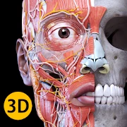
What is Anatomy 3D Atlas Apps?
Anatomy 3D Atlas Medical is a comprehensive interactive digital resource designed to present human anatomy with high-resolution three-dimensional models, detailed labeling, and layered anatomical systems. It offers users the ability to visualize bones, muscles, organs, nerves, and vasculature in isolation or in combination, facilitating spatial understanding of anatomical relationships. The interface typically supports rotation, zoom, cross-sectional viewing, and selective transparency to reveal underlying structures, making it valuable for illustrating depth and positional context. Built with meticulous morphological accuracy, the atlas often integrates descriptive text, clinical correlations, and functional annotations that translate static diagrams into dynamic learning experiences. Educational configurations may include quizzes, bookmarks, note-taking features, and customizable views that allow learners to tailor study sessions to specific topics or examination needs. Instructors can use the platform to create demonstrations and lecture materials, as the ability to highlight or hide structures streamlines explanations of complex regions. Medical professionals may find it useful as a quick visual reference when discussing anatomy with colleagues or explaining procedures to patients, because the realistic 3D reconstructions can improve comprehension compared with two-dimensional images. The product's architecture typically relies on optimized rendering engines to balance visual fidelity with responsive performance across different hardware profiles, and file formats are usually designed to facilitate rapid loading and smooth interaction. Support for multiple languages, measurement tools, and comparative anatomy modes enhances accessibility and utility for global audiences. Regular content updates and modular expansion allow the atlas to incorporate new discoveries and pedagogical improvements over time, keeping the resource relevant for both novice learners and experienced practitioners. Its integration with external educational systems and the capacity for exporting annotated images and customized lesson packages further strengthens its role as a versatile, scalable solution for modern anatomy education, research collaboration, simulation training, and continuing professional development across multiple healthcare disciplines worldwide.
As a pedagogical tool, Anatomy 3D Atlas Medical transforms traditional anatomy study by promoting active exploration and multisensory engagement. Students can manipulate anatomically accurate models to examine relationships between structures from arbitrary viewpoints, reinforcing three-dimensional thinking that often challenges learners who rely solely on textbooks or cadaveric dissection notes. Interactive labels and layered toggles help break down complex regions into manageable components, allowing progressive learning that aligns with curriculum sequencing. Integrated assessment modules support formative evaluation through timed quizzes, identification exercises, and spaced repetition features that promote long-term retention. Many implementations include customizable study paths whereby learners can focus on musculoskeletal anatomy for orthopedics, neuroanatomy for neurology, or regional anatomy for surgical preparation, optimizing study time and relevance. The ability to measure distances, angles, and cross-sectional areas aids students in correlating imaging slices with gross anatomy and understanding geometric relationships important for procedures and instrumentation. Annotation and note-taking tools let students capture personal insights, bookmark difficult areas, and compile personalized atlases for revision or group study. Collaborative functionalities can facilitate peer instruction and virtual lab sessions, enabling instructors to share prepared views, label sets, and guiding questions to structure interactive lessons. For learners with different accessibility needs, adjustable visual contrasts, scalable font sizes, and narrated descriptions provide alternative paths to understanding. When combined with problem-based learning scenarios, the atlas becomes a scaffold for clinical reasoning, helping students link structural knowledge with physiological function and pathological changes. By offering immediate, manipulable visual feedback, the resource accelerates correction of misconceptions and supports iterative learning cycles, thereby improving readiness for practical examinations, clinical rotations, and standardized testing. This practical, learner-centered approach complements gross anatomy courses and modern medical curricula that emphasize competency, integration, and lifelong learning skills. Frequent exposure to dynamic models shortens learning curves and builds confident spatial reasoning across clinical contexts.
Clinicians and allied health professionals can leverage Anatomy 3D Atlas Medical as a practical reference that complements imaging modalities and procedural planning. When interpreting radiological studies, the atlas's manipulable three-dimensional reconstructions and sectional views assist in correlating CT and MRI slices with gross anatomy, clarifying spatial relationships that affect diagnosis and intervention. Surgeons and interventionalists may use the tool to rehearse operative approaches virtually, exploring anatomical corridors, vascular branches, and neural pathways to anticipate critical landmarks and potential hazards. The atlas also supports preoperative education for patients by providing clear, realistic visuals that can demystify planned procedures and enhance informed consent discussions. Physical therapists, occupational therapists, and rehabilitation specialists benefit from detailed musculoskeletal layers that illustrate muscle origins, insertions, innervation, and lever arms, which aids in designing targeted exercise programs and understanding mechanics of injury. Emergency medicine and trauma teams can use the resource as a quick refresher on regional anatomy during acute care scenarios when rapid orientation is essential. In specialties such as dentistry, ENT, and ophthalmology, fine-grained models of craniofacial structures and orbital anatomy improve procedural planning and prosthetic fitting. The atlas's measurement and annotation tools facilitate documentation, interdisciplinary communication, and the creation of case-based teaching files that preserve important clinical findings. Integration with clinical workflows permits the export of labeled images and surgical plans to electronic records or educational materials, streamlining knowledge transfer. For research and simulation, the platform often supports customized model adjustments and data overlays, enabling experimental visualization of pathological states or device interactions. Throughout clinical practice, the atlas functions as a visual lingua franca that aligns terminology, reduces ambiguity, and strengthens collaborative decision-making among diverse care teams, thereby contributing to safer, more efficient patient management. Regular use cultivates anatomical fluency and supports evidence-based care by facilitating visual communication of complex anatomic concepts across specialties.
From a technical perspective, Anatomy 3D Atlas Medical combines high-fidelity anatomical modeling with performance-optimized rendering to deliver smooth, intuitive interaction on a range of devices. Models are constructed using detailed mesh geometry and texture mapping that capture surface contours, material properties, and subtle morphological variations between regions. Lighting models, ambient occlusion, and optional shading enhance depth perception, while level-of-detail algorithms reduce polygon counts dynamically to maintain responsiveness during rotation and zoom operations. Cross-sectional slicing and multiplanar reconstruction are implemented to align virtual anatomy with clinical imaging orientations, enabling side-by-side comparison and synchronized navigation between three-dimensional views and two-dimensional slices. The user interface typically emphasizes direct manipulation metaphors—pinch-to-zoom, drag-to-rotate, and tap-to-select—paired with contextual menus for layer visibility, labeling, and measurement tools, which streamline common workflows. Data formats support efficient loading of bundles that include model geometry, metadata, annotation layers, and localized text, facilitating modular content updates and language support. Export capabilities often allow generation of high-resolution screenshots, labeled diagrams, and structured reports for educational or clinical documentation. Accessibility considerations include keyboard navigation, screen reader compatibility for descriptive content, and adjustable rendering presets to accommodate different hardware capabilities. Security and privacy features focus on safe handling of user-created content and annotations, with local storage options and encryption for sensitive materials. Performance metrics are optimized by caching strategies, progressive loading, and GPU acceleration when available, minimizing latency in interactive sessions. Interoperability with common educational standards and file types promotes integration into learning management systems and institutional resources, while developer APIs may enable programmatic access to model views, annotations, and measurement data for custom research, simulation, or assessment tools. Regular profiling and updates to rendering pipelines maintain compatibility with evolving graphics drivers and operating environments, while modular content pipelines support addition of pathology libraries and instrument models to satisfy institutional teaching requirements and research.
No single tool perfectly captures every educational and clinical need, and Anatomy 3D Atlas Medical has inherent limitations and areas for improvement that users should understand. Despite high anatomical fidelity, models are abstractions that may not reflect individual patient variability, congenital anomalies, or rare pathological distortions; clinicians must integrate model-derived insight with patient-specific imaging and clinical context. Similarly, while 3D visualization enhances spatial comprehension, tactile experience from cadaveric dissection and hands-on procedural training remains indispensable for developing psychomotor skills, haptic feedback, and the subtleties of tissue planes. Networked collaboration features and multimedia integration can be extended to support more robust synchronous teaching, real-time annotation exchanges, and embedded formative assessments that capture learner performance analytics at scale. Interoperability improvements with clinical imaging platforms, simulation systems, and electronic health records could streamline the incorporation of de-identified, patient-specific models into preoperative planning and research while preserving privacy. Advances in volumetric rendering, physics-based tissue simulation, and augmented or virtual reality delivery will further close the gap between virtual practice and hands-on experience, enabling immersive rehearsals and instrument interaction with realistic force responses. Ongoing expansion of pathological atlases, procedural walkthroughs, and specialty modules will increase relevance across surgical, medical, and allied health disciplines, while multilingual content and culturally contextualized teaching materials can broaden accessibility. Scalability challenges persist when deploying large model libraries in bandwidth-limited or low-resource environments; offline caching strategies and lightweight model variants help mitigate these issues but require careful management. Finally, rigorous educational research and outcome studies are needed to quantify learning gains, transfer to clinical performance, and long-term retention attributable to 3D atlas use, guiding evidence-based adoption and continuous refinement of features. Stakeholder feedback loops, measurable competency benchmarks, and integration of simulated procedural metrics will accelerate iterative improvement and validate the pedagogical and clinical impact of three-dimensional anatomical learning tools globally relevant.